
NCRM in Ophthalmology
NCRM's first collaborative work in India started in the field of Vision. After several months of interaction, preliminary research work, exchange of information and exchange visits starting from 2002, we decided to take the cause of giving a cure to blindness.
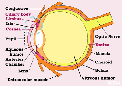
Fig.1 Anatomy of the eye
(Illustration not to proportions)
Human eye is a highly complex phenomenon, in which there are several mechanisms acting coherently to yield us the "Vision". Cornea is the transparent portion through which light rays pass in (Fig 1), which is controlled by the aperture of pupil with "Iris" and then the "Lens" focuses the incoming light signals on to the screen called "Retina" which is again one of the wonderful creations of the nature that converts the physical light into a neural signal perceivable by our higher optical centre in the brain and sends the signals through the "Optic Nerve". The vision could be jeopardized even any if one of the above components of the eye are damaged.
We have taken up cornea to start with because it is one components which is very much affected in our country especially among the socio economically underprivileged due to physical, and chemical injuries and infectious & persistent ulcers.
Cornea has five layers (Fig.2 ; quotes only three layers). The outer most layer is the epithelium which comes in to contact with the atmosphere. Cornea is an avascular (not supplied by blood vessels) component of the eye. Next layer is the stroma, beneath which the inner most endothelium is situated. The Red dot-encircled portion is the junction between the Cornea and the Sclera, which is called the Limbus and in this Limbus, the Corneal epithelial limbal stem cells are present. These stem cells when necessary, multiply and repair the damages of the corneal epithelial cell layer. When there is a damage to this limbus itself (Eg stevens-johnson syndrome) or when the repairing capacity of the limbal stem cells are over come by the disease process (Eg. persistent corneal ulcer), damage to the corneal epithelium results in opacity affecting the vision.

Fig.2 Corneo-Scleral junction; Limbus
(Illustration not to proportions)
Presently, almost all the publications in the literature report either the usage of 3T3 feeder layers (Mouse fibroblasts) or Human amniotic membrane in culturing the human corneal limbal stem cells. With our technology we have made it possible to culture the human limbal stem cells without using any biological materials either as nutrient feeder layer or as substrate, thus avoiding any human or animal protein in the tissue culture. With these developments we hope in the near future, repairing the corneal epithelial damages of any patient using his/her own health limbal stem cells, by culturing the same without amniotic membrane or feeder layers in the laboratory and applying them on the damaged portion of the eye will be possible.
Our aim is also to work on endothelial cell layer of the cornea as well as finding a solution to retinal diseases of both genetic and degenerative nature using cell therapeutics.